Fall 2003 Capstone Project: Radiation Therapy Optimization
This project comes from Our Lady of the Lake PET Imaging Center. The center is seeking to automates cancer detection and treatment into a more precise process.
Current methods of cancer detection do not use any mode of computer suggestion. Good methods of such computed intervention are being sought. Following the discovery of cancerous tissue in a patient, the process of treatment planning begins. The physician must find the best way to destroy the cancerous cells, often choosing the method of external radiation beam therapy. In this type of treatment gamma-ray beams are directed through the body at several angles to deliver a lethal amount of radiation to the cancer cells. By using many angles the dose given to healthy cells in kept to a minimum.
Imaging techniques at this point are much more advanced than the treatment techniques. Simply, the lasers used for treatment are much more crude than the cameras used for imaging. However, it is expected for engineers to come up with even more sophisticated treatment techniques and the objective here is to create mathematical models to govern the future machines. We use imaging information to optimize treatment. We use AccuImage software to extract the images and several Matlab toolboxes (such as wavelets, optimization, imaging processing) to create routines.
Our mapping strategy was to start by creating a simplified two-dimensional model of a slice matrix (similar to chat might be seen on PET or CT scans). From there, we tried to add more complexity to the model without substantially increasing the time it took for the program to run.
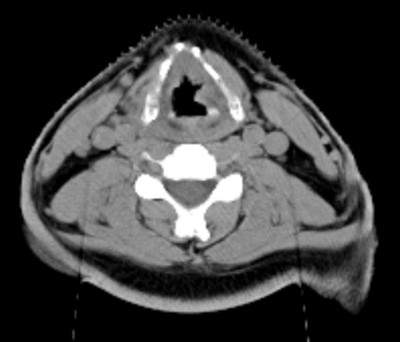 |
| CT scan shows show malignity in neck. However, it is impossible to determine exact position, size and shape of the tumor. |
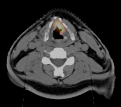 |
| Combined CT and PET scan shows tumor in neck. However, it is clearer where the cancer is, but it's not so easy to see what are the exact edges if it. |
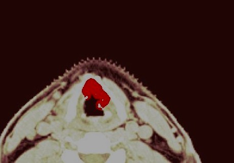 |
| Using the algorithm created at the Capstone clinic, it is possible to separate tumors in complete details. |
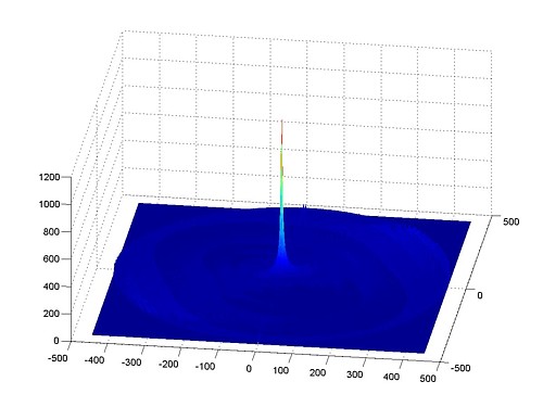 |
| Radiation beams are passed towards tumor through different angles in a large number of angles. Radiation accumulates in the "hot spot", center of tumor activity, while the healthy objects receive only low amounts of energy. |